Impaction of Maxillary Permanent incisor due to super numrary tooth is not a common entity encountered in dental practice but when present, it poses a disturbing esthetic dilemma to children and their parents. Early diagnosis and interception in these cases is the best way for their management. The purpose of this report is to describe the diagnosis and the clinical management of an impacted supernumerary tooth, which impeded the eruption of the permanent maxillary central incisor.
Keywords
Dentistry, Tooth, Treatment
Super numerary tooth, or hyper dontiais defined as a development a anomaly of number characterized by the presence of an extra tooth in addition when compared to the normal formula.1,2 The termmesiodens refers to a super numerary tooth located in the anterior region between the maxillary central incisors. It presents clinically the most frequent of all the super numerary teeth.3 A mesiodensis the most common cause of central incisor impaction, followed by odontomas and trauma.3
The exact etiology of mesiodens tooth remains unclear. Several hypotheses have been established for the formation of super numerary teeth which include egenetic and environmental factors, syndromic conditions and disturbances in dental development.4,5 A sex-linked pattern has also been suggested, as males are affected twice as frequently as females.3
The most common clinical complications of mesiodens include: central diastema, crowding, rotation, dislocation, delayed eruption of permanent incisor, abnormal tooth eruption, abnormal occlusion development, resorption of the roots of the adjacent incisors and cystic degeneration.3,6
The timing of the surgical removal of super numerary teeth remains highly controversial.1,2 Interceptive treatment has been advocated by some clinicians who believe that early extraction before radical formation of the permanent central incisor increases the chances of spontaneous eruption. Others have advocated delayed treatment or late removal after completion of the formation of the root of permanent incisor, to lower the risk of iatrogenic surgical damage to the permanent central’s apical development.7,8
The aim of this article was to present the diagnostic elements of super numerary tooth and the different therapeutic management.
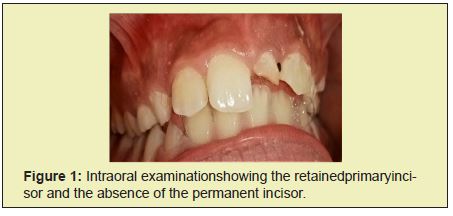
A 10-year-old girl presented to the Department of Pedodontics and Preventive Dentistry, at the Faculty of Dental Medicine of Monastir (Tunisia) with the chief complaint of unerupted left permanent maxillary central incisor (21) and the persistence of the deciduous tooth (61). The patient had no significant medical and family history. Extra oral examination revealed a convex facial profile and the presence of good facial balance in all proportions. Clinical examination revealed mixed dentition stage, retained deciduous left central incisor, missing permanent left central incisor and a midline shift towards the left (Figure 1). The permanent right central incisor, the right and the left lateral incisors and permanent first molars were fully erupted.


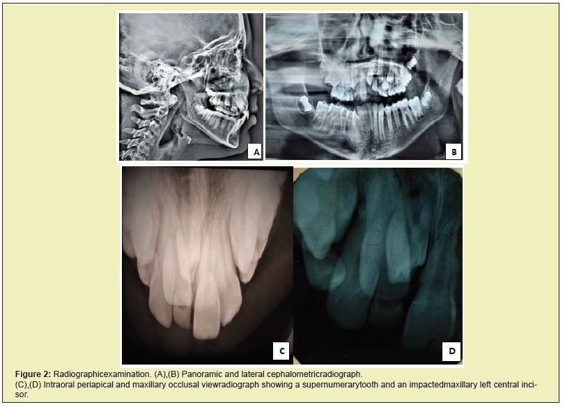
Radiographic examination which was done by panoramic, maxillary occlusal and retro alveolars radiographs showed the presence of a super numrary tooth which was present in the maxillary anterior region and an impacted left central incisor (Figure 2). Based on clinical and radiographic examination, the diagnosis made was the impaction of the upper left central incisor due to a super numrary tooth.
The treatment plan was the surgical removal of the super numerary tooth to initiate the eruption of the central incisor as its root was not fully formed.
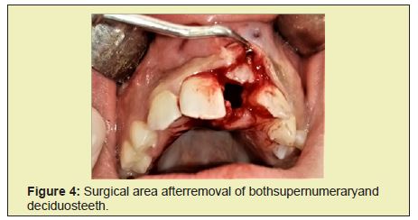
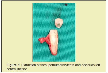
When the buccal flap was raised, the retained permanent incisor and the super numrary tooth were visible (Figure 3). The super numrary tooth and the temporary tooth were extracted (Figure 4 & Figure 5), and the flap was sutured (Figure 6).


The patient was recalled after 1 week for suture removal. Clinical and radiographic follow-ups were established to monitor the eruption of the upper left central incisor. The tooth erupted partially after 6 months (Figure 7) but the space required for proper alignment of the central incisor was insufficient, therefore, the girl was referred to Orthodontic Department.

Maxillary permanent incisors have a crucial part in facial esthetics and oral function.9 A delay in the time between the exfoliation of deciduous tooth and eruption of the permanent successor may be related to the disorder known as dental impaction. Impaction of these teeth has several adverse effects on smile and causes serious concerns and anxiety in child and his parents. It has been established that the main cause of the impaction of upper central incisors are super numerary teeth.10,11
In addition, physical barrier such as, over retained primary teeth, super numerary teeth ,tooth agenesis, tooth malformation or dilaceration, cysts or otherpatho logic obstruction in the eruptive path, insufficient space, developmental normalities, presence of a dense mucoperiosteum or sub mucosa,altered eruption sequence, trauma, palatal clefts, and genetic disorder, can act as etiologic factors. 9,11 The etiology of the occurrence of super numerary teeth remains unknown although several theories are proposed: the dichotomy theory, atavism (phylogenetic reversion) and hyperactivity of the dental lamina.8 A combination of genetic and environmental factors has been suggested.5
Morphologically the types of super numerations that occur include: supplemental tooth which refers to the super numerary teeth which are of normal shape and size called also incise form, rudimentary that describes a tooth with abnormal shape and smaller size which includesconical, tuberculate, molariform or odontomas, the latter being composite or complex.12-14 It may occur singularly, multiplarly and unilaterally or bi laterally.12 A supernumerary tooth should be suspected when there is a symmetry in the eruption pattern of the maxillary incisors.15
Diagnosis occurs between seven and nine years of age, probably because of delayed eruption of the permanent central incisors, thus intraoral periapical, maxillary occlusal and/or panoramic radiographs are the key at this stage to conclude if the unerupted tooth is absent, included, or impacted.16 The treatment of impacted central incisor in children is challenging and the ideal timing of surgical removal of a supernumerary tooth is subject to controversy2: immediate versus delayed intervention. Among the problems and risks of immediate intervention are potential damage to adjacent teeth resulting in devitalisation and/or root malformation of adjacent teeth, and the fact that the young patient is unable to psychologically tolerate the surgical procedure. In the other hand, The complications related with untreated super numerary teeth include: crowding, loss of space, loss of eruptive force, occlusal interference, midline shift malocclusion over retention of primary teeth, delayed eruption of permanent incisors, impaction, midline diastema, axial rotation or inclination of erupted permanent incisors, root resorption or dilacerations and pulp necrosis.11,13,17 Less common sequel included large follicular sacs, cystic degeneration and nasal eruption.
The treatment modality for impacted permanent incisors is: surgical extraction of impacted supernumerary tooth and observation till the spontaneous eruption of permanent incisor that may occur when the root is still developing. Once the root apex has closed, the tooth loses its potential to erupt.16,17 In the present case since the root was not completely formed it was desirable to wait for spontaneous eruption. Orthodontic traction of the impacted incisor, when the apex was almost closed, by means of either removable or fixed appliance in cases where the ectopically erupted or unerupted incisors need assistance to be brought into the right position.14
The average time of the spontaneous eruption of impacted maxillary central incisors varies from 3 to 30 months.16,11 Theoretically the factors that may be responsible for the failure of eruption of the permanent upper incisors after the elimination of the obstructive super numerary teeth are: the degree of apical displacement, the maintenance of sufficient arch space, the chrono logical age, the degree of root maturity, the initial location and axial inclination of the impacted tooth, the root curvature and its degree of formation of the impacted tooth, the scar formation due to the operation, the removal of part of the gubernaculums long which teeth would erupt; and conical as opposed to trabeculated form of the super numerary tooth.18
When the incisors do not erupt at the expected time it is crucial to determine the exactetiology and formulate an appropriate treatment plan. Early diagnosis prevents complications associated with supernumerary tooth and its based essentially on clinical and radiographic examination.
None.
None.
Author declares that there is no conflict of interest.
- 1. Wen-Yu Shih, Chun-Yi Hsieh, Tzong-Ping Tsai. Clinical evaluation of the timing of mesiodens removal. J Chin Med Assoc. 2016;79(6):345–350.
- 2. Primosch RE. Anterior supernumerary teeth--assessment and surgical intervention in children. Pediatr Dent. 1981;3(2):204–215.
- 3. Kocatas Ersin N, Candan U, Riza Alpoz A, et al. Mesiodens in primary, mixed and permanent dentitions: a clinical and radiographic study. J Clin Pediatr Dent. 2004;28(4):295–298.
- 4. Rehan Qamar Ch, Javed Bajwa, Muhammad Imran Rahbar. Mesiodensetiology, prevalence, diagnosis and management. POJ. 2013;5(2):73–76.
- 5. Van Buggenhout G, Bailleul-Forestier I. Mesiodens. Eur J Med Genet. 2008;51(2):178–181.
- 6. Hong J, Lee DG, Park K. Retrospective analysis of the factors influencing mesiodentes eruption. International Journal of Paediatric Dentistry. 2009;19(5):343–348.
- 7. Ayers E, Kennedy D, Wiebe C. Clinical recommendations for management of mesiodens and unerupted permanent maxillary central incisors. European Archives of Paediatric Dentistry. 2014;15(6):421–428.
- 8. Van Buggenhout G, Bailleul-Forestier I. Mesiodens. Eur J Med Genet. 2008;51(2):178–181.
- 9. Noorollahian S, Shirban F. Chair time saving method for treatment of an impacted maxillary central incisor with 15-month follow-up. Dent Res J (Isfahan). 2018;15(2):150–154.
- 10. Gupta M, Pandit IK, Gugnani N, et al. Management of Impacted Maxillary Central Incisor with Surgical Exposure and Orthodontic Traction-Two Case Reports. Indian Journal of Dental Sciences. 2013;5(3): 67–69.
- 11. Jain M, Namdev R, Bodh M, et al. Multidisciplinary management of an impacted permanent central incisor associated with two unerupted supernumerary teeth: A case report. 2014;8(6):21.
- 12. Leyland L, Batra P, Wong F, et al. A retrospective evaluation of the eruption of impacted permanent incisors after extraction of supernumerary teeth. J Clin Pediatr Dent. 2006;30(3):225–231.
- 13. Ata-Ali F, Ata-Ali J, Penarrocha-Oltra D, et al. Prevalence, etiology, diagnosis, treatment and complications of supernumerary teeth. J Clin Exp Dent. 2014;6(4):e414–e418.
- 14. Cogulu D, Yetkiner E, Akay C. et al. Multidisciplinary Management and Long-Term Follow-up of Mesiodens: A Case Report. J Clin Pediatr Dent. 2008;33(1):63–66.
- 15. Kathleen Russel A, Magdalena Folwarczna A. Mesiodens-Diagnosis and Management of a Common Supernumerary Tooth. J Can Dent Assoc. 2003;69(6):362–366.
- 16. Cosme-Silva L, Costa LL, Silva M. Et al. Combined Surgical Removal of a Supernumerary Tooth and Orthodontic Traction of an Impacted Maxillary Central Incisor. J Dent Child. 2016;83(3):167–172.
- 17. Rallan M, Rallan NS, Goswami M, et al. Surgical management of multiple supernumerary teeth and an impacted maxillary permanent central incisor. BMJ Case Rep. 2013:bcr2013009995.
- 18. Witsenburg B, Boering G. Eruption of impacted permanent upper incisors after removal of of supernumerary teeth. Int J Oral Surg. 1981;10(6):423–431.

