Tissue expander is a plastic surgery that is of great advantage to regenerated aesthetics for the patient because the skin flap is near the injury as well as it can reduce scar at the donor and receive site. But the method requires 2 stages of surgery, a long process of pumping the sac, the cooperation of the patients and their family, especially the close monitoring of medical staff to bring about optimal results. When the expansion sac is exposed at the end of the pumping procedure, the patient should be surgically removed the bag and the lesion should be immediately covered, to avoid the shrink of expanded skin which cause the reduction of covered size of lesion. This clinical case is a 13-year-old female with a large hairless scalp area in size 16x19cm and 2 expander bags which were fully pumped and some necrosis in the expanded skin flap. Additional, she unfortunately got hepatitis by Cytomegalo virus (CMV), Epstein-Barr virus (EBV). Treatment include treating hepatitis, removing expander bags, but the second surgery was delayed 2 weeks because she has the severe hepatitis; as a result, only 50% of the lesion is covered.
Keywords: Tissue expander, Skin, Scalp, Hair loss
Abbreviations: CMV: Cytomegalo virus; EBV: Epstein Barr virus
Tissue expander is a natural phenomenon that has been written in the medical literature for a long time such as laxity of abdominal wall, expansion of cranium during pregnancy. Tissue dilation can also be seen in diseases such as obesity, tissue expansion around benign or malignant tumors. It can also be seen in cases of placing ear rings, stretching lips or stretching the neck with necklaces in African and Asian tribes. In 1950, Neumann first applied this technique by using a rubber ball to make auriculaplasty.1,2
The main principle of this method is to place beneath the healthy skin near the lesion with one or several expandable bags made of silicone, then slowly inflate with isotonic saline through the valve. The bag is inflated upon selected volume then remove the bag, remove the lesion and transfer the flap. This technique allows creating new tissue that has almost completely same color and sense.1,2
At Trung Vuong Hospital of Vietnam, this technique has been carried out by Pham Trinh Quoc Khanh since 2002. The process of tissue expansion is performed in 2 stages. Stage 1 is to put a tissue expansion bag and pump the bag for the first time about 10-20% of the bag volume, after choosing a bag of suitable shape and size. After placing the bag 1-2 weeks, start pumping, once a week, about 10-15% of the bag volume each time, but must ensure capillary perfusion is good. Stage 2 lasts about 7 days, the patient was admitted to the hospital to remove the tissue expansion bag, resect the lesion and tranfer the expanded tissue to cover defective tissue and complete the reconstruction.3-7
In comparison with other plastic surgery techniques such as skin grafting, pedicle flaps, free flaps, etc…, tissue expander can limit scarring in donor site as well as receive site which has highly aesthetic due to using the skin tissue which is nearby the damaged skin.5
Indications
Locations where can be applied tissue expansion techniques compose of skull, face, neck, trunk, extremities in order to treat burn scars that cause skin contracture in the elbow, groin, neck, and perineum: of removing melanoma, tattoo: of reconstructing mammary tumorectomy; in regenerating the hairy scalp due to accidental loss of scalp and baldness but still has hair area on the scalp.1-3
Physiology
When the mechanical pressure on the skin increases, there will be 2 phenomena of mechanical and biological dilation. Cellular morphological changes occur in response to tension. Mechanical dilation due to cell tension, bio dilation is the proliferation of cells due to disruption of cleft junctions and increased tissue surface area. The epidermis is thicker; the dermis is thinner with collagen binding. This effect is maximal after 6-12 weeks of implantation. At the molecular level, it is a variety of cytokines that are produced in response to tissue dilation.1,2
The blood vessels in the tissue dilation area increase in number and size. Neoangiogenic factors such as vascular endothelial growth factor will be more expressed.1,2
Major complications include
Infection at the area of implantation, open incision, exposure of the expander sac, ischemia and tissue necrosis, damage to the expander sac, hematoma and seroma. However, complications are preventable and treatable.8
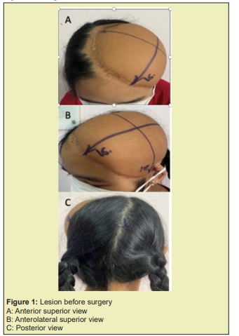
A 13-year-old female patient, with a history of losing her hairy scalp 6 years ago due to being pulled her hair into a motorboat engine, then she underwent surgery to cover the loss of the scalp with a free anterior lateral thigh flap. This flap area is about 16x19cm in size on the top-temporal (Left) area without hair, affecting aesthetics negatively, making children feel self-deprecating. No other disease was noted Figure 1.
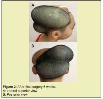
The patient had first surgery on September 28th, 2022 when 2 tissue expansion sacs were placed: sac 1 with size 6x18cm, V=600cc, in the temporal region (R), sac 2 with size 9x12cm, V=700cc, in the occipital area Figure 2.
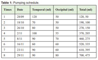
The patient expansion sacs were pumped with normal saline. Pumping schedule is listed in the Table:
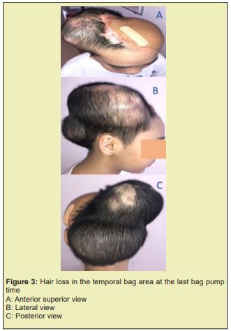
In the times of sac pumping, we always checked the skin condition before pumping, the capillary perfusion test was made after each the bag pump times, and the last 3 times there was hair loss in the temporal bag area Figure 3.
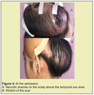
On December 8th (9 weeks after sac placement) Patient was admitted to the hospital with a stretch of the scar that exposed the sac, necrotic anemia at 2 corner of size #3x4cm on the scalp above the temporal sac area. The patient declared that she had worn a hat hugging tightly her head every school day for 1 week before admission.
Clinical examination
Dilated surgical scar, skin tear in 2cm long, dry necrotic anemia 2 protruding corners in size #3x4cm on the scalp above the temporal sac area, opaque exudate through the skin tear. When we palpate dilating area, it felt flabby. Vital signs are normal, no fever Figure 4.
The preoperative examination showed elevated liver enzymes AST 73, ALT 154, and white blood cell 22.56, Neu 77.4%, Pro calcitonin 0.061<0.1. Other tests were within normal limits.
Treatment
The patient was removed the tissue expander sac 1 day after admission, cleaned the compartment beneath the scalp, inserted anti-adhesive gauze into this compartment. Antibiotic using follows the antibiogram. Consultation with infectious doctor about hepatitis of unknown cause, immunoassay showed that patient was infected with 2 types of CMV and EBV virus simultaneously (CMV IgM Positive 206.2AU/ml, EBV VCA IgM Positive 19.8, EBV VCA IgG Positive 42.5U/mL).
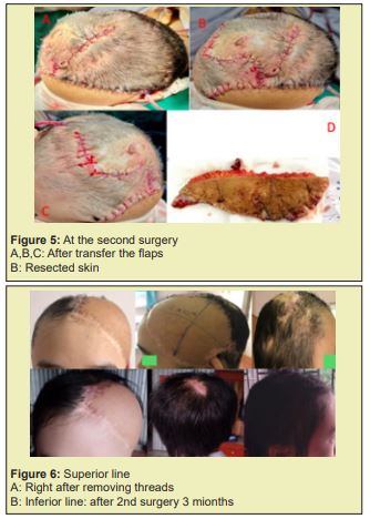
After treating hepatitis, the patient was surgically removed part of the hairless skin flap at the top-left temporal and transferred the flap from the area of tissue expansion. The scalp flap after removing the sac 2 weeks ago was constricted and 2 necrotic 3x4cm removal flaps, making it difficult to rotate the flap in order to cover lesion and reducing the area amount of the covered lesion. Blood lose #300ml. The patient was transfused with 1 unit of red blood cells Figure 5.
After 2 weeks the wounds healed completely, the hairless flap was reduced in area from 16x19cm to 10x15cm (#50% area reduction) Figure 6.
Indications to cover the skull after craniofacial skin peel 6 years ago with a free flap showed technical success but did not regenerate the patient's hairy area. In this patient, skin grafting will be gentler because it also achieved the goal of covering the damage to regenerate the hairy skull area afterwards.
Tissue expander is a technique indicated to regenerate hairless scalp effectively, which gain high aesthetic results when the scalp with hair is stretched and moved to cover the hairless scalp. Although hair follicles may be sparse, this is still a more effective method than hair transplantation.3 In this clinical case of a very large hairless scalp area of 16x19 cm, which affects aesthetics and low self-esteem of 13-year-old child - at puberty, it is appropriate to indicate intervention by tissue expander technique.
2 bags were placed at the right temporal (700cc) and occipital (475cc) areas suitable for the goal of covering all of the entire hairless scalp area.
The patient ’s expander sac had been inflated for 8 weeks, reached the desired volume as required, the sac has been inflated to full volume, the area of expanded skin is enough to cover the hairless skull. She was scheduled to admission for surgery after 1 week to remove the bag, remove the lesion, and transfer the expanded skin flap to cover the hairless skull area.
But when the patient was admitted to the hospital, the bag was exposed about 2cm in diameter at the temple. With the complication of exposed bag when the bag was inflated; normally, the patient was surgically removed the bag and immediately used a stretched skin flap to cover the lesion; but unfortunately, this patient was jaundiced and was found to have hepatitis due to CMV and EBV at the time of bag exposure. The second surgery required general anesthesia, so that the patient was delayed for 2 weeks for stable treatment of viral hepatitis. During hepatitis treatment, the patient was bandaged and used gauze to reduce the retraction of the expanded skin. However, the skin flap was still shrinking and could only cover about 50% of the hairless area.
In the duration of inflating the sac, the patient is far away, so it is difficult to monitor and examine her after the sac is pumped. Besides that, the patient often uses a hat hugging tightly the area of the tissue expansion, especially the circumference of the bag, the corners. In addition, during the pumping process, although the capillary perfusion problem is checked, it is inevitable that the pump area may be too tense. These problems can be related to ischemic complications and partial necrosis of the skin in the expanded area, scar dilation and sacs exposure, and infection of sac compartment. In the last 3 times of bag pumping, the patient appeared some areas of hair loss in the temporal region; this is a sign that the skin is dilated with anemia. However, blood circulation in the dilated skin with hair loss is still satisfactory 1-2" after pressing the finger. After the last pump, the patient was not closely monitored and the patient wore a cap that tightly pressed the scalp, increasing tissue ischemia and led to necrosis of a part of expanded skin and exposure of the expander sac.
Tissue expander is a plastic surgery that has good advantage of restoring aesthetics for the patient because of the skin tissue in the vicinity of the injury. But the method requires 2 stages of surgery, a long process of pumping the sac, the cooperation of the patients and their family, as well as the close monitoring of medical staff to bring about optimal results. When the expansion sac is exposed at the end of the pumping progress, the patient should be surgically removed bag and the lesion should be immediately covered, to avoid shrinkage as in this patient's case, which cause reduction in the covered size of lesion.
None.
None.
The author declares no conflicts of interest regarding the publication of this paper.
- 1. Ivo Alexander Pestana, Louis C Argenta, Malcolm W Marks. Principles and Applications of Tissue Expansion. Plastic Surgery–Principles,Elsevier Inc. 2018:pp.473-497.
- 2. Janae L Maher, Raman C Mahabir, Joshua A Lemmon. Tissue Expansion. Essentials of Plastic Surgery. Quality Medical Publishing Inc. 2014:pp.57-66.
- 3. Guilherme J Christiano, Nicholas Bastidas, Howard N Langstein. Reconstruction of the Scalp, Calvarium and Forehead, Grabb and Smith’s Plastic Surgery, Lippncott Williams and Wilkins. pp.342-351.
- 4. Jason E Leedy, Smita R Ramanadham, Jeffrey E Janis. Scalp and Calvarian Reconstruction, Essentials of Plastic Surgery. Quality Medical Publishing Inc. 2014pp.382-391.
- 5. Jeffrey E Janis, Bridget Harrison. General Management of Complex Wounds. Essentials of Plastic Surgery. Quality Medical Publishing Inc. 2014:pp.10-16.
- 6. Phạm Trịnh Quốc Khanh. Nhận xét kỹ thuật vạt da giãn trong phẫu thuật tạo hình tại Bệnh viện Cấp cứu Trưng Vương, Tạp chí Y học thảm hoạ và Bỏng. 2005;3:52-63.
- 7. Trần Thiết Sơn. Vạt tổ chức giãn, Phẫu Thuật Tạo Hình, Nhà xuất bản Y học. 2005:pp.67-71.
- 8. Cunha MS, Nakamoto HA, Herson MR, et al. Tissue expander complications in plastic surgery: a 10-year experience. Rev Hosp Clin Fac Med Sao Paulo. 2002;57(3):93-97.

