Cutaneous squamous cell carcinoma (SCC) of the nipple areolar complex (NAC) is a rare entity with only a few cases reported in the medical literature. The standard treatment for invasive squamous cell carcinoma includes complete surgical excision of the lesion with postoperative assessment of free margins. Although this skin cancer involves the NAC it should be considered as a completely separate entity from other breast cancers, and therefore may be treated differently than breast cancers involving the NAC. Mohs micrographic surgery is a well-accepted technique for non-melanoma skin cancers (NMSC) with its remarkable precision for tumor eradication and tissue-sparing properties which favor excellent aesthetic outcome. We present a clinical case of 75-year-old Caucasian female who presented to our clinic with a papulose, scaly lesion located at her right nipple that was confirmed to be SCC of the nipple. The patient underwent complete excision of the tumor using Mohs micrographic technique with immediate reconstruction of spared NAC using purse-string suture technique. Free surgical margins were determined intra-operatively by Mohs’s cryosurgery method and further assessed by permanent evaluation. Follow-up was assessed for 3 years without evidence of local recurrence, and high patient satisfaction with the final aesthetic outcome. To the best of our knowledge, this is the first report that validates the feasibility of application of the Mohs technique for nipple SCC excision with immediate nipple reconstruction. Our results suggest that Mohs micrographic surgical technique may be a safe treatment modality for SCC lesions of the NAC allowing superior tissue preservation and enhanced aesthetic results compared to standard wide local excision.
Keywords: Mohs micrographic surgery, Nipple, Squamous cell carcinoma
Breast cancer is the most common malignancy encountered in woman world-wide. Histologically, invasive breast cancers are commonly derived from ductal or lobular origin, with invasive tumors from stromal origin being rarer. Since multiple breast ducts terminate at the nipple, the nipple areolar complex (NAC) may be commonly be involved form breast cancers located at or near it. Invasion of the skin of the NAC by the primary breast malignancy may also occur and require special surgical attention as part of the surgical treatment for breast cancer. In these cases, wide local excision with complete removal of the NAC may be needed to achieve safe surgical margins. Squamous cell carcinoma (SCC) of the nipple however, is a completely separate malignant entity, and is not part of the breast malignancies.1 Therefore, the treatment of SCC of the NAC should not be dictated from the treatment required for breast cancer. Since this uncommon cancer does not originate from the breast tissue it should be treated in a distinct manner and thus complete removal of the NAC may be unjustified and even viewed as overtreatment.
Mohs micrographic surgery (MMS) is a well-accepted technique for non-melanoma skin cancers (NMSC) with a 99% precision rate for tumor eradication and tissue-sparing properties providing excellent tumor control and aesthetic outcome.2 Here we present a case of a 75-year-old woman with SCC of the nipple that was successfully treated using Mohs micrographic surgical technique. To the best of our knowledge, this is the first report that validates the application of the Mohs technique for nipple tumor excision with intra-operative assessment of tissue margins and immediate nipple reconstruction. We present the following case in accordance with the CARE reporting checklist.
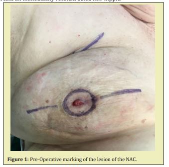
A 75-year-old Caucasian female presented to our clinic with a papulose, scaly lesion located at her right nipple. Her past medical history was significant for right breast ductal carcinoma in-situ (DCIS) that was treated with lumpectomy followed by adjuvant radiotherapy. Other comorbidities included diabetes mellitus, hypothyroidism, and atrial fibrillation. Lesion biopsy confirmed squamous cell carcinoma of the nipple. Mammography and breast ultrasound were normal. The patient underwent excision of the tumor using Mohs micrographic technique Figure 1. Excision was commenced until confirmation of clear margins during surgery, followed by wound closure using purse-string technique in layers to create an immediately reconstructed neo-nipple.
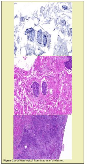
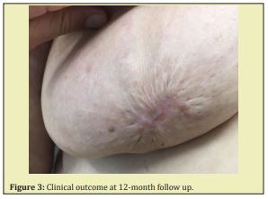
The total excised tissue measured 35*35*20mm and samples were submitted for complete and final histological examination. The examination revealed a 15mm diameter ulcerated well differentiated squamous cell carcinoma of the nipple Figure 2. The tumor was connected to the epidermis and infiltrated through the whole thickness of the dermis and the upper part of the subcutaneous fat. There was no evidence of ductal breast tissue or intravascular invasion of the tumor cells. The tumor cells stained positively for p63 and were negative for CK7. The histological morphology of the tumor, its location (skin and the upper part of the subcutaneous fat without involving the underlying breast tissue), and the immunophenotype of p63+/CK7- supported the diagnosis of SCC originating from the skin. At 12-month follow-up there were no signs of local recurrence, and the patient was well content with the final aesthetic outcome and declined the opportunity of nipple-areolar tattooing Figure 3. Similarly, during the 3 year follow-up period no signs of local recurrence occurred or changes in aesthetic outcome.
All procedures performed in studies involving human participants were in accordance with the ethical standards of the institutional and with the Helsinki Declaration (as revised in 2013). Written informed consent was obtained from the patient.


SCC is the second most common form of skin cancer,3 with 1 million cases diagnosed every year in the United States.4 Cutaneous SCC of the nipple and areolar skin however is a rare entity with only a few cases reported in the medical literature.1,5 Surgical treatment options for invasive squamous cell carcinoma include standard wide surgical excision with postoperative margin assessment. The value of Mohs technique in other parts of the skin body have been well established for the treatment of NMSCs showing precise excision of tumor and enabling nearly 100 percent of microscopic control rates of the surgical margin.2 This technique allows precise removal of the tumor with minimal amount of uninvolved surrounding tissue. Compared to other techniques used, the complete marginal visualization in Mohs technique decreases the need for re-interventions and allows excellent cure rates with maximal preservation of unaffected tissue. This in turn enables a large diversity of immediate reconstructive options and decreases the rates of deformity.6,7 Treatment of cutaneous SCC with MMS has shown similar if not superior cure rates compared to wide local excision (WLE), with cure rates up to 99% and 95% in MMS and WLE, respectively.2,8 Since the majority of cutaneous malignancies do not invade past the epidermis and dermis to other organs, the use of Mohs micrographic surgical technique for SCC of the nipple may provide a good alternative to WLE, preserve the underlying breast tissue and facilitate the use of different flaps and grafts.
This case demonstrates the use of Mohs surgical technique in a patient with cutaneous SCC of the nipple who may have otherwise undergone a standard approach of extensive wide excision of the NAC and underlying breast tissue resulting in significant changes in breast contour. The 3-year follow-up results presented here suggest that Mohs surgical technique may be feasible and safe alternative for the treatment of SCC lesions. Since the NAC and underlying breast tissue could be partially spared after accurate margin analysis to prevent recurrences, the shape and volume of the breast were preserved minimizing defect size.Moreover, we suggest that the purse-string closure technique should also be considered as a simple technique resulting in increased areolar convexity and reduction in loss of nipple projection, promoting good aesthetic result. This preliminary successful experience suggests that further studies should be performed to define the role of Mohs technique as the treatment of choice for SCC of the nipple.
None.
The authors have no financial interest to declare in relation to the content of this article.
Author declares that there is no conflict of interest.
- 1. Dye K, Saucedo M, Raju D, et al. A common cancer in an uncommon location: A case report of squamous cell carcinoma of the nipple. Int J Surg Case Rep. 2017;36:94–97.
- 2. Tolkachjov SN, Brodland DG, Coldiron BM, et al. Understanding Mohs Micrographic Surgery: A Review and Practical Guide for the Nondermatologist. Mayo Clinic Proceedings. 2017;92:1261–1271.
- 3. Karia PS, Han J, Schmults CD. Cutaneous squamous cell carcinoma: Estimated incidence of disease, nodal metastasis, and deaths from disease in the United States, 2012. J Am Acad Dermatol. 2013; 68(6):957–966.
- 4. Rogers HW, Weinstock MA, Feldman SR, et al. Incidence estimate of nonmelanoma skin cancer (keratinocyte carcinomas) in the US population, 2012. JAMA Dermatol. 2015;15:1081–1086.
- 5. Pendse AA, O’Connor SM. Primary Invasive Squamous Cell Carcinoma of the Nipple. Case Rep Pathol. 2015;327487.
- 6. Buker JL, Amonette RA. Micrographic surgery. Clin Dermatol. 1992;10:309–315.
- 7. Hanke CW. Frederic Mohs Tribute. History of Mohs micrographic surgery. J Drugs Dermatol. 2002;1(2):169–174.
- 8. Alcalay J. The value of Mohs surgery for the treatment of nonmelanoma skin cancers. J Cutan Aesthet Surg. 2012;5(1):1–5.

