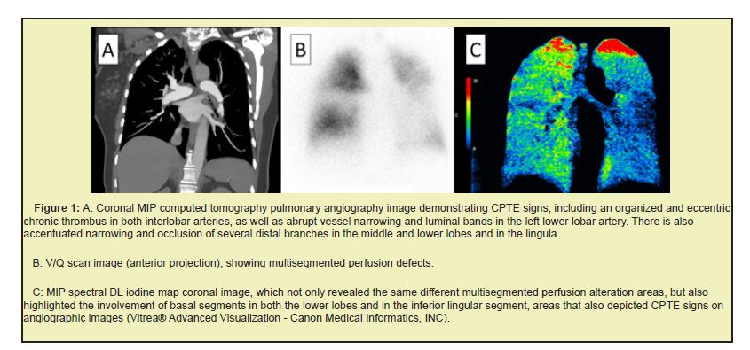
Pulmonary scintigraphy is considered a technique of choice in the evaluation of chronic pulmonary thromboembolism. Technical advancements in computed tomography as dual-energy (spectral) have allowed obtaining high quality images. As an example, a computed tomography as dual-energy patient case is shown, who underwent both scintigraphy and spectral deep learning not only was a lower radiation dose observed scintigraphy but also provided anatomical data on perfusion defect, essential for surgical planning and also evidenced other areas of perfusion alteration that were not clearly identified on V/Q scintigraphy. Thus, we bring this initial experience with the use of Spectral Deep Learning as a possibility to combine anatomical and perfusional assessment in the same acquisition, reducing dose and possible costs.
Keywords: Tomography, Pulmonary, Clinical research
V/Q scan is considered the technique of choice in the evaluation of chronic pulmonary thromboembolism (CPTE).1 In the last decade, technological advances in computed tomography (CT) have allowed for the acquisition of high-quality images while using lower levels of radiation, allowing for the detection of small thrombus in segmental and subsegmental regions.2 Although some studies demonstrated higher sensitivity in V/Q scan, more recent studies have showed better sensitivity in CT.1
The term dual-energy CT (DECT) or spectral CT, refers to the tomographic acquisition that uses two different energy spectra providing both morphological and functional information of the lung parenchyma, which contributes to the process of the pathology assessment.2,3 However, most of the available DECT technologies have limitations in the FOV and others in the use of automatic exposure control (AEC). The Aquilion ONE/PRISM Edition equipment-Canon Otawara, Japan DECT, is a deep learning (DL) reconstruction algorithm that allows the ultra-fast switching of kVp (80-135), with volumetric anatomical coverage, routine use of the AEC and 50cm FOV on acquisition.
Spectral DL reconstruction is based on the large amount of information contained in the low kVp and high kVp data, which are iteratively compared generating high quality images. Deep Learning Views (DLVs) are generated by the trained neural network using measured data from both the opposite energy views as well as adjacent same energy views. The use of DLVs results in highly precise temporal and spatial alignment helping to ensure accurate material classification and minimal temporal artifact.4,5
DECT has several clinical applications, among them, the iodine map, which allows for the visualization, analysis and quantification of the iodine distribution. Iodine, air, and soft tissue are the three most frequently analyzed materials in the thorax. With DECT, the attenuation of iodine is exceedingly greater at 80Kvp than at 135Kvp. Thus, DECT may improve detection of pulmonary embolism in comparison with conventional CT and assist in the evaluation of lung perfusion.6 As an example, a CPTE patient case is shown, who underwent both V/Q scan and spectral DL Figure 1.

Spectral DL not only had a lower radiation dose (V/Q scan: 7.55 mSv and spectral DL: 6.72 mSv), but also provided simultaneously the anatomical data on perfusion defect, essential for surgical planning.2,3,6
The results confirmed and showed excellent agreement with the perfusion defects seen on V/Q scan and evidenced other areas of perfusion alteration that were not clearly identified on V/Q scan but also demonstrated correlation with segmental e subsegmental arterial branches with signs of CPTE seen on computed tomography pulmonary angiography images. As a result, we introduce this preliminary experience with the use of Spectral Deep Learning as a possibility to combine anatomical and perfusion assessment in the same acquisition, potentially reducing dose and costs.
None.
This Short Communication received no external funding.
Regarding the publication of this article, the author declares that he has no conflicts of interest.

